
A different Approach to Patient Care
You Make the Decision.
Our Responsibility Is to Help You Understand Your Options
Choosing surgery for a child is a major decision.
There may be several reasonable approaches, including hearing devices, observation, or surgery. Each option has potential benefits, limitations, and risks.
Dr. Kesser believes that families should have the information they need to make an informed decision.
Our role is to evaluate, explain, and guide.
The decision belongs to you and your family.

What is Aural Atresia?
Aural atresia is condition where a baby is born without an ear canal. The eardrum may not have formed properly or may be missing, and there can also be issues with the middle ear bones (ossicles). However, the inner ear and auditory nerve are usually unaffected. Most babies with aural atresia also have microtia, which is a small or misshapen outer ear.
Aural atresia leads to hearing loss, but with appropriate treatments, most children can experience improved hearing.
Congenital Aural Atresia
Congenital aural atresia is a condition in which the ear canal does not fully develop before birth. The eardrum and middle ear structures may also be underdeveloped to varying degrees. Aural atresia can affect one ear or both ears and is frequently associated with microtia, an underdeveloped or malformed outer ear.
How Does Aural Atresia Affect Hearing?
The outer and middle ear normally work together to collect sound and conduct it toward the inner ear. When the ear canal is absent or severely underdeveloped, sound cannot travel normally to the eardrum and middle ear.
As a result, most patients with aural atresia have conductive hearing loss. Importantly, the inner ear is often normally developed, which means the structures responsible for sensing sound may still function normally.
Unilateral and Bilateral Aural Atresia
Aural atresia may occur on one side (unilateral atresia) or on both sides (bilateral atresia).
Children with unilateral atresia may have difficulty hearing speech in background noise and may have difficulty determining where a sound is coming from. Research involving congenital unilateral aural atresia has examined the benefits of restoring hearing to the affected ear, particularly for hearing in noisy environments and binaural hearing.
For children with bilateral atresia, early hearing assessment and appropriate hearing support are particularly important while the child's anatomy and treatment options are being evaluated.
Can Aural Atresia Be Repaired?
In selected patients, atresia repair surgery can create a functional ear canal and restore a pathway for sound to reach the middle and inner ear.
The operation may involve creating the ear canal, freeing the hearing bones, constructing a new eardrum, and lining the reconstructed canal with a skin graft. Whether surgery is appropriate depends on the individual anatomy of the ear, including the middle ear, hearing bones, facial nerve, and surrounding structures.
Every Patient Is Different
Aural atresia is not the same in every child. The anatomy of the middle ear and temporal bone can vary significantly from patient to patient.
A careful evaluation may include:
Hearing testing
Physical examination
High-resolution temporal bone CT
Assessment of the middle ear and hearing bones
Evaluation of the facial nerve and surrounding anatomy
These findings help determine whether a child may be a candidate for atresia repair and help guide surgical planning.
If your child has congenital aural atresia, understanding the anatomy is the first step toward understanding the available hearing options.
The Ear Canal
The ear canal, also called the external auditory canal, is the passage that carries sound from the outside of the ear toward the eardrum.
In a normally developed ear, sound travels through the ear canal and reaches the eardrum. Vibrations are then transmitted through the middle ear and hearing bones before reaching the inner ear.
What Happens in Aural Atresia?
In congenital aural atresia, the ear canal does not develop normally. In complete atresia, the ear canal may be absent, while in stenosis the canal is present but abnormally narrow. The eardrum and middle ear may also have varying degrees of developmental abnormality.
This difference is important because simply creating an opening on the outside of the ear does not necessarily create a functional hearing pathway.
Creating a Functional Ear Canal
Atresia repair is designed to create a new pathway through the temporal bone to the middle ear.
During surgery, the surgeon may need to:
Create an opening through the bone to form the new ear canal.
Identify and preserve the important surrounding structures.
Free the hearing bones when appropriate.
Construct a new eardrum.
Line the new canal with a thin skin graft.
Create and align the external opening, or meatus, with the reconstructed canal.
The goal is not simply to create a visible opening. The goal is to create a stable, skin-lined ear canal that allows sound to reach the middle ear and inner ear efficiently.
Why Anatomy Matters
The anatomy surrounding the ear canal is different in every patient.
Important structures considered during surgical planning include:
The middle ear space
The malleus, incus, and stapes
The oval window
The facial nerve
The temporal bone
The inner ear
A high-resolution temporal bone CT can provide important information about these structures and help determine whether atresia repair is anatomically appropriate.
Ear Canal and Long-Term Care
After atresia repair, the reconstructed canal requires careful follow-up. Healing, canal narrowing, debris accumulation, and other changes may need to be monitored over time.
Long-term hearing and canal health are important parts of the treatment process—not simply the day of surgery.
A successful atresia repair begins with understanding the anatomy and continues with careful long-term follow-up.
Conductive Hearing Loss
Conductive hearing loss occurs when sound cannot travel efficiently through the outer or middle ear to reach the inner ear.
In congenital aural atresia, the absence or underdevelopment of the ear canal and associated middle ear structures can prevent sound from being conducted normally toward the inner ear.
The Three Parts of Hearing
Hearing depends on the coordinated function of three major areas:
Outer Ear
Collects sound and directs it into the ear canal.
Middle Ear
The eardrum and three tiny hearing bones—the malleus, incus, and stapes—transmit and amplify sound vibrations.
Inner Ear
The cochlea converts sound vibrations into electrical signals that are sent to the brain.
In many children with aural atresia, the inner ear is normally developed even though the outer and middle ear have developmental differences. This is why the hearing loss associated with atresia is typically conductive rather than sensorineural.
Unilateral Conductive Hearing Loss
When atresia affects only one ear, the child may hear relatively well in quiet environments because the other ear is functioning normally.
However, having one normally hearing ear does not necessarily mean that hearing is normal in all situations.
Children with unilateral hearing loss may have difficulty:
Understanding speech in background noise
Hearing in a busy classroom
Following conversations when several people are speaking
Determining where a sound is coming from
Research involving children with unilateral congenital aural atresia has demonstrated measurable changes in hearing in noise following surgical correction.
Bilateral Conductive Hearing Loss
When both ears are affected, hearing access can be significantly reduced.
Early hearing evaluation and appropriate amplification or other hearing support can provide access to sound while the child's anatomy and long-term treatment plan are being evaluated.
How Is Conductive Hearing Loss Evaluated?
Evaluation may include:
Behavioral hearing testing
Bone-conduction testing
Auditory brainstem response testing when appropriate
Audiometry
Physical examination
Temporal bone imaging
These tests help distinguish conductive hearing loss from sensorineural hearing loss and provide important information for treatment planning.
Can Conductive Hearing Loss Be Improved?
The treatment depends on its cause.
For children with congenital aural atresia, options may include hearing devices, observation in selected circumstances, or surgical reconstruction of the ear canal when the anatomy is appropriate.
Atresia surgery is a highly anatomy-dependent procedure. Imaging and hearing evaluation are important parts of determining whether surgery may provide a meaningful hearing benefit. Research from the University of Virginia has also shown that preoperative anatomical factors, including middle ear development, can be associated with postoperative hearing outcomes.
The goal is not simply to improve a hearing test. It is to help the patient access sound more effectively in everyday life.
Specialty
Signs & symptorns
Aural atresia can occur in one or both ears, but it most commonly affects just one ear. When it affects only one ear, hearing in the other ear is typically normal. Since the ear canal hasn’t formed, there is no opening in the affected ear.
Cholesteatoma
A cholesteatoma is an abnormal growth behind the eardrum, in the middle ear, where small bones transmit sound from the eardrum to the inner ear. As the cholesteatoma grows, it can damage these bones, potentially causing hearing loss if left untreated. When you feel pressure or pain in or behind the ear or ringing in the ear, please go see a doctor as soon as possible.
Unknown
Doctors are not exactly sure why aural atresia occurs, but it is sometimes linked to genetic mutations. In some cases, children with aural atresia may also have a genetic syndrome, such as Treacher Collins syndrome or Goldenhar syndrome.
Diagnosed
If a baby has issues with the formation of their outer ear or fails the newborn hearing screen, doctors will conduct an exam and additional hearing tests to check for other concerns.
Imaging studies, like a CT scan, are typically not performed until the child is closer to 6 years old, as the temporal bone around the ear undergoes significant growth in early childhood.
How is aural atresia treated?
Babies with aural atresia require a care team to ensure they receive the best treatment. This team typically includes:
An otolaryngologist (ENT specialist) to plan treatment and perform surgery
An audiologist (hearing specialist) to address hearing loss
A speech therapist to assist with language development
A pediatric plastic surgeon to reconstruct the ear (if the child has microtia)
A pediatrician to coordinate care and manage any ear infections promptly
A geneticist to help families understand the hereditary aspects of aural atresia
Not every child with aural atresia will require surgery. Treatment depends on the formation of the middle and inner ear and the growth of the temporal bone during early childhood. For most children, a hearing device can significantly improve hearing.
How Do Doctors Trest Hearing Loss in Aural Atresia before they can receive surgical treatment?
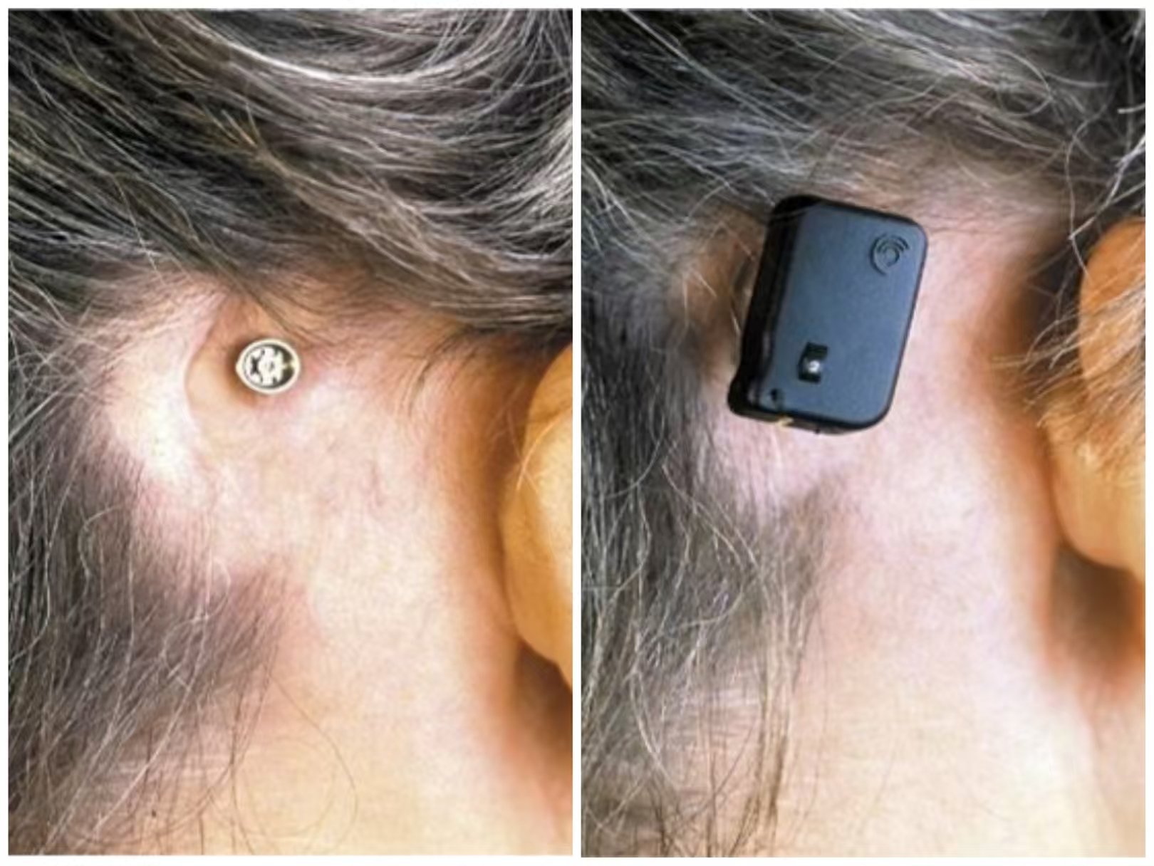
It’s important for a baby’s development to treat any hearing loss as soon as possible. Most children with aural atresia have a normal hearing nerve, so they can use a bone conduction device. This device bypasses the missing ear canal and takes sound vibrations directly to the hearing nerve.
On babies, the device usually is attached with an elastic band that goes around the child’s head. As kids grow, other hearing devices can help. Some can be surgically placed in the bone behind the ear or inside the ear.
Treatment
For patients with bilateral atresia, a bone conduction hearing device is definitely needed as early as possible. For those with unilateral atresia, the bone conduction hearing device is optional.
Once the patient reaches 4 years of age, they can come in for an evaluation.
Both surgery and the use of a hearing aid are reasonable options to consider.
Atresia repair surgery is one potential treatment, but it's important to note that the surgery cannot restore hearing to the normal range. It can only bring the hearing closer to normal levels.