
What Happens After Surgery?
Long-Term Follow-up Matters
Atresia repair does not end when the patient leaves the operating room.
During recovery, the reconstructed ear canal requires careful protection and monitoring. Infection, moisture, tissue growth, narrowing, and other problems can affect the long-term result.
Families receive postoperative instructions and ongoing guidance regarding:
Ear canal care
Cleaning
Water precautions
Hearing monitoring
Follow-up examinations
Warning signs
Local physician communication
Because patients often return to their home countries after surgery communication between the surgical team and local physicians can be especially important.
Three Types of Postoperative Rehabilitation Programs
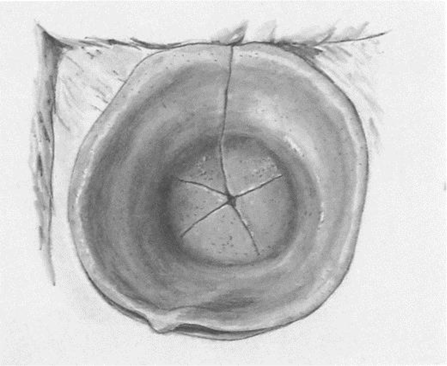
Regular Postoperative Rehabilitation
Regular postoperative rehabilitation refers to the care and follow-up activities that all patients are required to complete at scheduled time points after surgery, according to their surgeon's instructions. For example: on the day of surgery, one week, one month, three months, six months, and one year after surgery.

Personalized Postoperative Rehabilitation
Personalized postoperative rehabilitation refers to guidance tailored to each patient's individual recovery progress and the impact of surgery on their return to daily activities. Examples include recommendations on when it is safe to fly, swim, shower, resume sports and physical activities, or undergo surgery involving the outer ear.
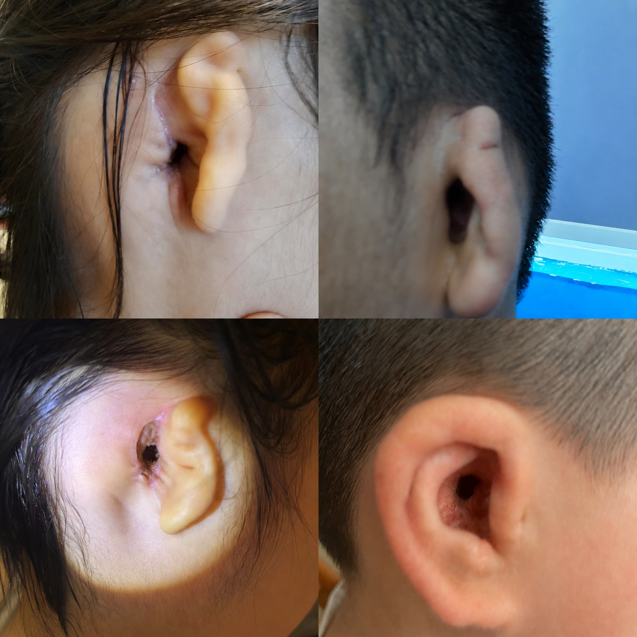
Postoperative Complication Management and Rehabilitation
This category addresses postoperative complications that may occur during the recovery period due to various causes. These may include redness and swelling of the ear canal, scar management, infection, unpleasant odor, eardrum perforation, or a sudden decline in hearing.
Routine Postoperative Rehabilitation
External auditory canal reconstruction is a delicate surgical procedure designed to restore the structure of the ear canal and improve hearing. Recovery follows a standardized timeline, with specific medical guidelines for postoperative care and follow-up at each stage. Strict adherence to your surgeon's instructions—including scheduled follow-up visits, wound care, and ear canal cleaning—helps reduce the risk of complications such as canal stenosis, debris or crust buildup, and infection, while promoting the long-term stability and function of the reconstructed ear canal.
A. Early Postoperative Period
Early postoperative period (dressing protection phase, 0–7 days):
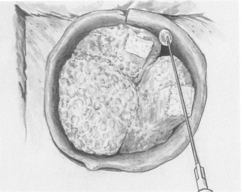
During surgery, a medical expandable sponge is placed according to the size of the ear canal defect to support the newly reconstructed canal, and a sterile dressing is applied externally to stabilize the surgical site.
Key postoperative care guidance:
The dressing must be fully protected at all times. Patients must not remove, adjust, or prematurely detach it. It should remain in place until it naturally loosens or is removed at the appropriate healing stage. Any external displacement may damage the newly formed ear canal flap and significantly increase the risk of canal collapse or scar overgrowth.
At the 7-day postoperative visit, the outer layer of the ear canal packing sponge will be removed by the physician, and the wound will be examined to assess healing status and check for signs of inflammation such as redness, swelling, or discharge.
B. Mid Postoperative Period
Full Packing Removal Phase (1 Month Postoperative)
The expandable sponge used during surgery is layered and placed in segments. While the outer layer is typically removed at the 7-day follow-up, deeper structural packing materials remain in place for approximately one month after surgery.
Key clinical information for this stage:
At the 30-day postoperative visit, the physician will completely remove all internal ear canal packing materials. This allows the first comprehensive examination of the reconstructed ear canal, confirming the basic structural formation of the new canal and evaluating the viability and condition of the surgical flaps.
C. Wound Healing and Repair Phase After Surgery
Two-stage professional ear canal cleaning (3 months and 6 months postoperatively)
The newly reconstructed ear canal is a surgically created structure. During the healing process, it continuously produces epithelial debris and crusting. If these materials accumulate, they may obstruct the ear canal and lead to itching or inflammation.
Therefore, two scheduled professional cleanings are required at approximately 3 months and 6 months after surgery to carefully remove debris and maintain canal patency.
3 Months Postoperative: Basic Examination and Cleaning
The primary purpose of this follow-up visit is a comprehensive evaluation of the deep ear canal to confirm that no residual packing materials remain after the 1-month removal stage. At the same time, surface crusting and accumulated debris are carefully removed to keep the ear canal clear and prevent long-term retention of secretions or blockage.
6 Months Postoperative: Advanced Deep Cleaning
At this stage, the ear canal enters a more stable healing phase. Crusts and older epithelial layers become more firmly adherent. Standard saline-based softening methods, which are suitable for natural ear canals, are often not appropriate for reconstructed ear canals.
Clinical note: Differences in softening agents for reconstructed ear canals
In normal healthy ears, cerumen is typically softened using saline irrigation. However, in reconstructed ear canals, the mucosal lining is more delicate and crusts are often dry and hardened.
Medical-grade baby oil provides stronger lubricating and penetrating properties, allowing better softening of hardened crusts. This reduces the risk of mucosal injury or bleeding during cleaning and significantly improves both patient comfort and procedural safety.
D. Long-term Maintenance and Stability Period (Up to 3 Years Postoperative)
The ear canal requires a long period to fully stabilize and for the mucosal barrier to become firmly established. The first three years after surgery represent a high-risk observation period for potential complications. It is recommended to establish a regular follow-up schedule, typically every six months, to monitor long-term healing and maintain ear canal stability.
Significance of Long-term Follow-up
Every 6 months, patients should undergo deep ear canal cleaning and structural evaluation. This allows the physician to detect early signs of scar hyperplasia, ear canal stenosis, or abnormal crust accumulation, and to intervene promptly before these issues progress.
If symptoms such as ear fullness, pain, discharge, or hearing decline occur during recovery, patients do not need to wait for the scheduled follow-up and should contact the Kesser surgical team immediately for timely management.
Rehabilitation after ear canal reconstruction is a staged, long-term systematic process. It includes four essential components: maintaining dressing position, staged removal of packing materials, phased ear canal cleaning, and long-term scheduled follow-up. Each step is designed according to the physiological healing process of the ear canal mucosa.
Patients should never skip follow-up visits or attempt self-management based on subjective feelings, as standardized postoperative care is critical to ensuring optimal long-term surgical outcomes.
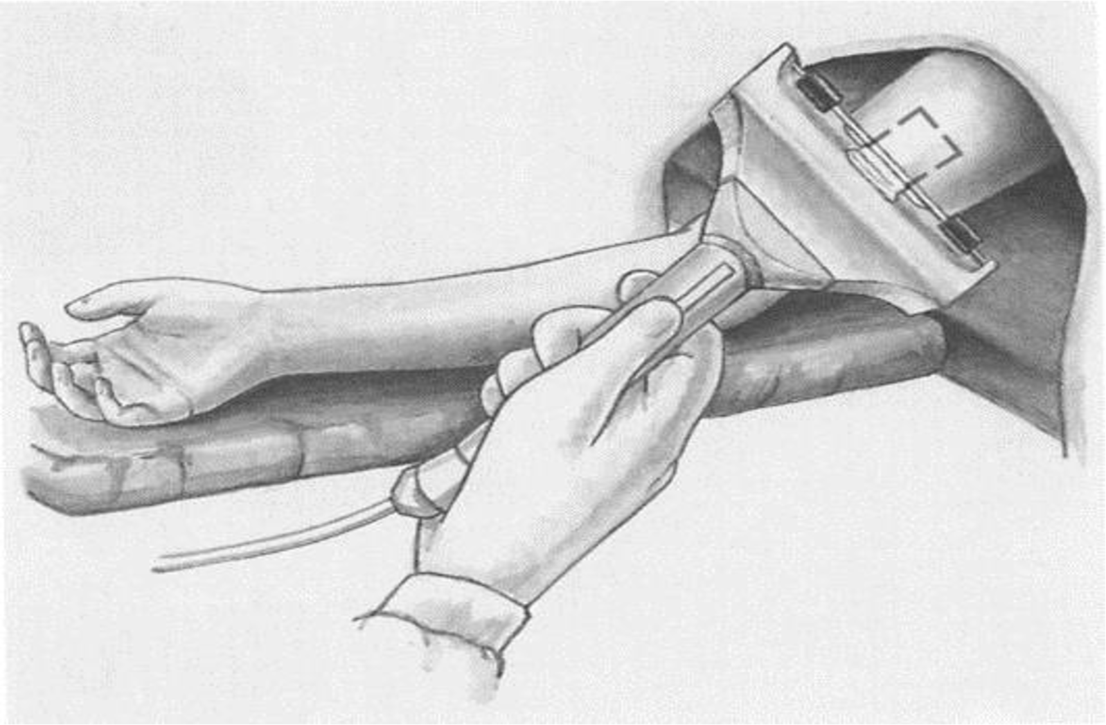
Regular Rehabilitation Plan
Postoperative rehabilitation is a critical component in achieving long-term successful outcomes after ear canal reconstruction surgery. To ensure proper healing of the surgical site and reduce the risk of complications, each patient should strictly follow the rehabilitation plan developed by the physician and complete follow-up visits and care at scheduled time points.
Typically, postoperative follow-up is required at the following intervals:
Day of surgery
1 week postoperatively
1 month postoperatively
3 months postoperatively
6 months postoperatively
1 year postoperatively
Rehabilitation after ear canal reconstruction mainly involves two areas:
The newly reconstructed ear canal
The skin graft donor/recipient area
Day of Surgery
After surgery, the physician will apply a firm dressing to the surgical area to protect both the newly reconstructed ear canal and the skin graft site. Therefore, patients generally do not need to worry about external impact on the wound.
It is especially important that the Telfa dressing covering the skin graft area remains in the correct position at all times. In the early postoperative period, even minor bleeding may cause the dressing to shift, which can affect graft adherence and increase the risk of scar formation or poor healing.
If the dressing becomes noticeably loose or partially detached, do not remove the Telfa dressing under any circumstances.
1 Week Postoperative
At one week after surgery, the physician will remove the outermost dressing and assess wound healing. This includes evaluating the survival of the skin graft and the recovery status of the reconstructed ear canal.
1 Month Postoperative
At approximately one month after surgery, the physician will remove the packing materials used to support the newly reconstructed ear canal.
Because the transplanted skin inside the ear canal continues to undergo healing and gradually forms keratinized debris and crusts, ongoing monitoring and care are still required in the subsequent recovery period.
3 Months and 6 Months Postoperative
As the newly reconstructed ear canal gradually matures, crusts, keratin debris, and secretions may accumulate inside the canal. It is recommended that at 3 months and 6 months after surgery, patients undergo professional cleaning performed by an experienced otology specialist under a surgical microscope. This helps maintain canal patency, supports normal epithelial growth, and reduces the risk of ear canal stenosis and infection.
1 Year Postoperative
At one year after surgery, a comprehensive follow-up is performed to evaluate the long-term stability of the ear canal, hearing recovery, and the maturity of the ear canal epithelium. If recovery is satisfactory, the patient may transition to routine annual follow-up care.
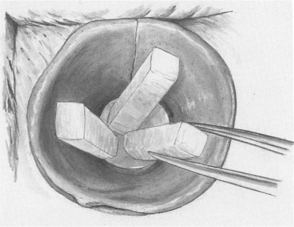
After artificial ear canal reconstruction surgery, there is no universal timeline for returning to daily activities, travel, or sports. Recovery must be individualized based on each patient’s wound healing progress and the development status of the reconstructed ear canal.
Changes in air pressure and exposure to humid or aquatic environments can irritate the newly formed, fragile ear canal and the repaired eardrum. Inappropriate activities may lead to complications such as pain, infection, or deformation of the ear canal.
Personalized Rehabilitation
Effects of Air Pressure Changes and Safe Timing for Flying After Surgery
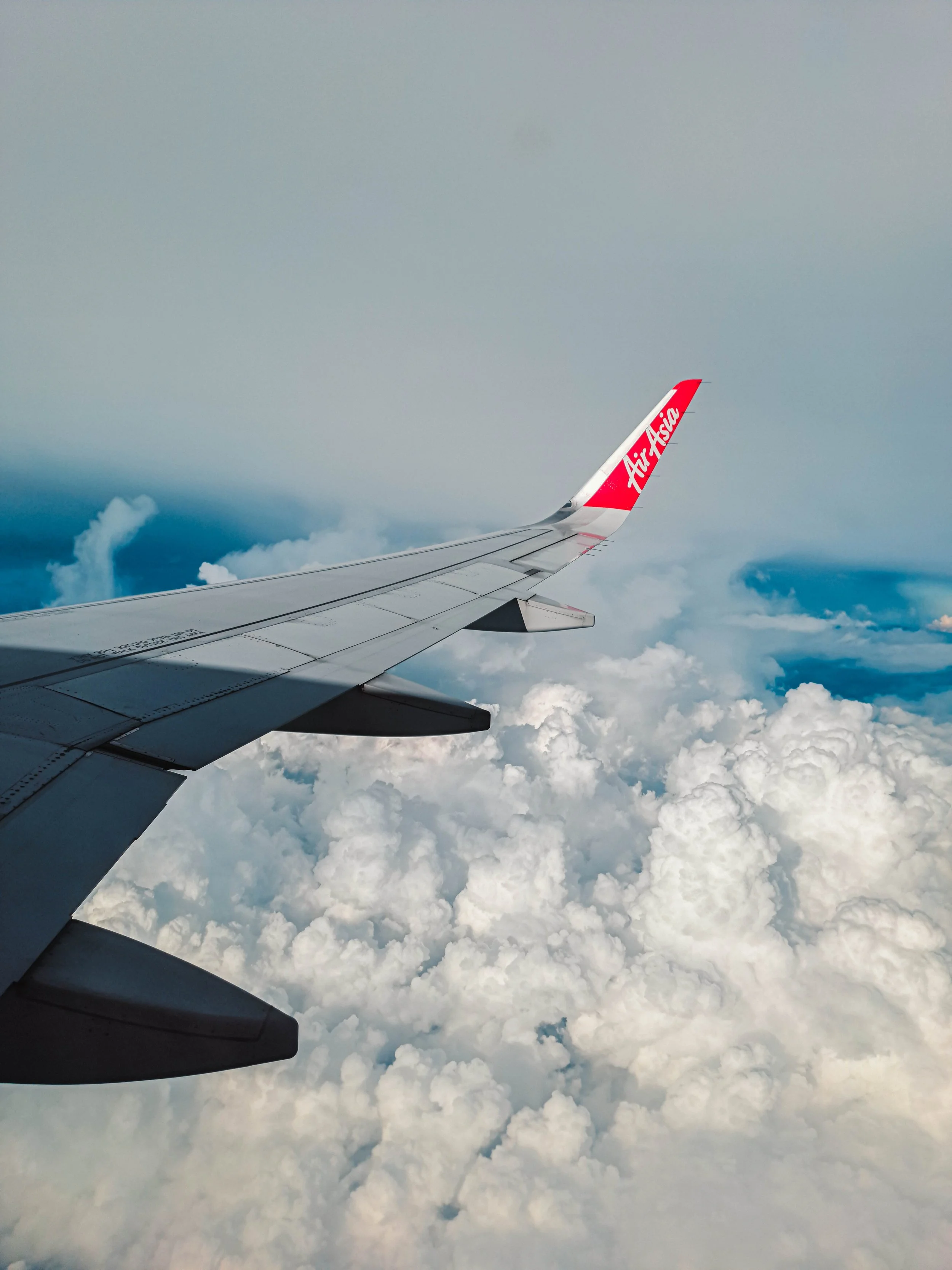
During aircraft takeoff and landing, significant pressure differences occur between the inside and outside of the eardrum. If the newly reconstructed ear canal and repaired eardrum have not yet fully stabilized, these pressure changes may cause tissue congestion, displacement, or delayed healing.
Recovery adaptation period
Clinical recommendations suggest that the ability to safely fly should be evaluated individually between 1 to 4 months after surgery. Early travel is not recommended without medical assessment.Optimal safe timing
Around 4 months postoperatively is generally considered a safer milestone for air travel. By this time, the newly formed ear canal has typically developed sufficient structural thickness and stability to better withstand pressure fluctuations during flight.Medical advice
If urgent travel is necessary, patients should undergo a clinical review in advance. The physician must evaluate healing status before determining whether short-distance flying is safe.
— Air Travel Considerations After Surgery

Distinguishing Routine Exercise from High-Risk Water Activities
Most routine land-based exercises such as running and strength training do not cause damage to a reconstructed ear canal. Patients only need to avoid direct trauma or impact to the ear area.
In contrast, water-related activities are considered high-risk during recovery and should be strictly avoided. The risks mainly come from two sources:
Chemical exposure in swimming pools
Chlorine and other disinfectants used in artificial water systems are highly irritating. The newly reconstructed ear canal mucosa is delicate, and prolonged exposure may lead to redness, itching, chronic inflammation, and disruption of epithelial healing.Microbial risk in natural water environments
Lakes, rivers, and seawater contain large amounts of bacteria, microorganisms, and parasites. Water entry into the ear canal can easily cause infection and, in severe cases, may damage the reconstructed canal structure.High-risk patient group
Patients within 10 months after surgery, as well as pediatric patients, should strictly avoid all humid water environments, including swimming, diving, and water sports.Long-term protection strategy (after full healing)
Even after full recovery is confirmed, patients engaging in water activities should maintain ear dryness and cleanliness. Protective ear drops designed for swimmers may be used to form a protective barrier on the ear canal surface, reducing the risk of dermatitis and proliferative complications.
— Sports
Petroleum Jelly Simple Protection Method
Water entering the ear during bathing is one of the most common triggers of postoperative inflammation. Even small amounts of contaminated water can soften crusts and promote bacterial growth if exposure is prolonged.
A simple and effective waterproofing method is recommended:
Before showering, apply a sufficient amount of petroleum jelly evenly around the entrance of the external ear canal. Petroleum jelly has excellent water-repellent properties, helping divert water away from the ear canal opening and significantly reducing the risk of water entering the reconstructed canal. This helps maintain a dry internal environment and lowers the risk of infection.
Summary for patient education
Reconstruction of the artificial ear canal involves a long-term process of mucosal remodeling. Factors such as air pressure changes and moisture exposure can continuously influence long-term recovery outcomes.
Return-to-activity timelines for travel, exercise, and bathing must be individualized according to each patient’s recovery progress. Patients should not follow others’ recovery schedules.
If symptoms such as ear fullness, pain, or discharge occur, patients should promptly contact the Prof. Kesser surgical team for evaluation and management.
— Daily Shower Waterproofing
Common Postoperative Complications and Personalized Rehabilitation Guidance After Ear Canal Surgery
During the postoperative rehabilitation process after ear canal surgery, some patients may experience mild abnormal symptoms. It is important to first understand that these symptoms are not always directly related to the surgery itself. For example, common illnesses such as colds or fever may lead to secondary conditions like otitis media, which can also cause ear canal redness, swelling, or middle ear fluid accumulation.
Even if symptoms such as ear redness, discharge, odor, eardrum perforation, recurrent stenosis, or hearing decline occur, this does not necessarily mean there will be permanent damage to hearing.
Management of these conditions should be individualized based on the patient’s actual recovery status and otoscopic examination findings. The following sections provide an overview of the causes of common complications and their corresponding tailored treatment approaches.
1. Infectious Complications: Redness, Discharge, and Ear Odor
1. Infectious complications: redness, discharge, and ear odor
After ear canal surgery, symptoms such as redness, fluid discharge, or unpleasant odor are often related to wound infection, inadequate postoperative care, or accumulation of secretions within the ear canal. Management should always be based on otoscopic (endoscopic ear examination) findings to avoid improper handling that may worsen the condition.
Ear redness:
This is commonly caused by mild inflammatory irritation of the surgical wound. After confirming the infection status via otoscopy, appropriate antibacterial ear drops may be prescribed to reduce inflammation and relieve redness while protecting the newly formed tissue.
Ear discharge (oozing):
Postoperative exudation is a relatively common finding during healing. Under the guidance of the surgical team, specially prepared medicated powder may be used to help control discharge and promote a dry healing environment for the surgical site.
Ear odor:
Odor is usually not an isolated symptom and often occurs together with discharge. Accumulated exudate, dead skin, and external debris can remain in the ear canal, creating an environment for bacterial growth and odor formation. Professional cleaning of the ear canal is required first, followed by standardized topical treatment to fully resolve both odor and underlying infection.
2. Ear Canal Stenosis: Staged Personalized Management Plan
2. Complication of Ear Canal Stenosis: Staged Personalized Management
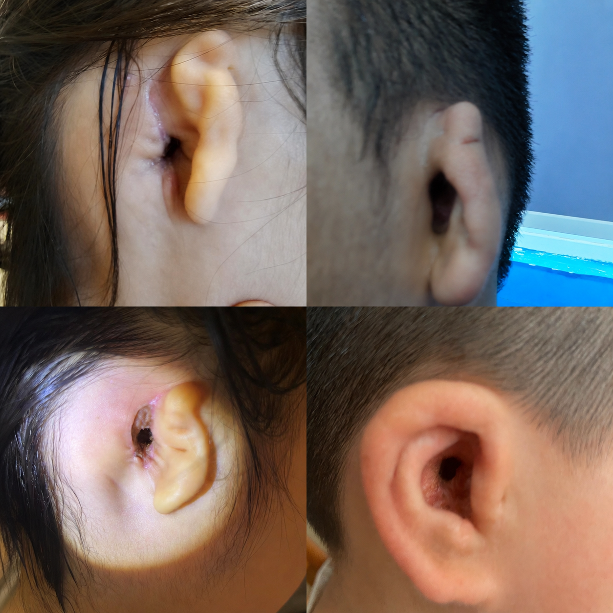
Ear canal stenosis is a key postoperative complication that requires careful prevention and monitoring. It is mainly caused by tissue overgrowth at the ear canal opening or contraction of the healing wound. Clinical observation suggests that narrowing of the ear canal most commonly occurs during nighttime rest, while the canal remains relatively stable during the daytime.
A stepwise, individualized treatment approach is applied depending on severity:
Mild early stenosis:
This stage is characterized by initial narrowing or early contraction of the ear canal. It is particularly common in pediatric patients. During sleep, a dilating ear stent may be worn, combined with anti-proliferative medication to effectively suppress tissue contraction and maintain normal canal shape.
Moderate progressive stenosis:
If early intervention is insufficient and narrowing continues, the dilating stent may be replaced with medical-grade expandable sponge. In combination with antibacterial ear drops, the sponge expands to support the ear canal, prevent further stenosis, and reduce infection risk.
Severe proliferative stenosis:
In cases of significant narrowing with obvious tissue hyperplasia, targeted injections such as lidocaine or specialized steroid medications may be used to strongly inhibit scar formation and tissue overgrowth, thereby halting progression of stenosis. Clinical data indicate that this intervention is effective in controlling severe cases, with no reported cases of uncontrollable progression.
3. Important Educational Notice
All symptoms of complications, treatment approaches, and rehabilitation protocols described in this article are based on clinical individualized experience and are not universally applicable.
Each patient’s surgical wound, baseline ear canal condition, and healing capacity differ. The severity of complications, timing of intervention, and treatment outcomes will therefore vary from person to person.
If any abnormal symptoms occur after surgery, the underlying cause must first be confirmed through otoscopic examination. A personalized rehabilitation plan should then be developed by a qualified physician. Patients should not self-manage based on symptom comparison, as improper handling may negatively affect overall postoperative recovery outcomes.